While X-ray is not the only way to measure a scoliosis curve, it is today’s gold standard. That being said, as X-rays are taking an image of a 2-D space and scoliosis is a 3-D condition, X-ray results should be combined with other measurements and observations for a comprehensive understanding of the condition.
4 Key Takeaways
- The Role of X-Rays in Scoliosis Diagnosis: X-rays are crucial for diagnosing scoliosis, providing a reliable, accessible, and cost-effective method to measure spinal curvature and determine treatment paths with the Cobb angle measurement as a key diagnostic tool.
- Scoliosis and Growth Patterns: Understanding a patient’s growth pattern is essential in predicting scoliosis progression, especially in adolescents. Monitoring growth through standing and seated X-rays helps tailor treatment to slow or halt curvature progression during critical growth phases.
- Limitations of Scoliosis X-Rays: While X-rays are invaluable, they have limitations due to their 2-D nature, representing a 3-D condition. Comprehensive understanding requires combining X-ray data with physical examinations and other assessments for a complete treatment strategy.
- Importance of Specialized Scoliosis Treatment: Expert interpretation of scoliosis X-rays and a multidimensional treatment approach are crucial for effective scoliosis management. Specialists use 3-D considerations to develop customized, patient-centered treatment plans beyond what X-rays alone can offer.
What a Scoliosis X-Ray can Tell Us
One aspect of scoliosis that people get confused with is that just because it’s not preventable doesn’t mean it’s not treatable. In fact, inaction is the worst thing a person can do, or not do when it comes to a scoliosis diagnosis.
Here at the Scoliosis Reduction Center®, we customize each and every treatment plan to the individual needs and abilities of our patients. The first step before designing an effective treatment plan is getting thorough measurements and assessments that give a full picture of the individual, the curvature, where the curvature is located, degree of flexibility, and the Cobb angle measurement. The gold standard for measuring a spinal curvature is the scoliosis X-ray. It is crucial to include imaging of the lumbar spine, along with other spinal sections, to accurately assess scoliosis and classify the specific type of curvature.
What is a Scoliosis X-Ray?
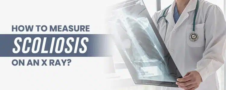
A scoliosis X-ray is a diagnostic imaging test that plays a pivotal role in evaluating the curvature of the spine in individuals with scoliosis. This essential tool is used not only for diagnosing scoliosis but also for monitoring its progression and assessing the effectiveness of treatment. By providing a two-dimensional image of the spine, a scoliosis X-ray allows doctors to measure the degree of curvature and identify any potential issues. This imaging technique is invaluable in giving a clear picture of the spine’s alignment and helping to guide treatment decisions.
Why X-Rays are so Important for People with Scoliosis
Discovered in 1895, you might think that since then, a more sophisticated form of technology than the X-ray would have been developed. Today, X-rays are still the most reliable method for assessing and measuring a spinal curvature. In addition to their reliability, they are quick, accessible, and inexpensive.
As the most common way to obtain a Cobb angle measurement, X-rays have stood the test of time. A Cobb angle measurement designates a patient’s curvature as mild, moderate, or severe; this is determined by the Cobb angle measurement that assesses how far the scoliosis spine bends and twists away from a straight alignment.
If a spinal curvature measures greater than 10 degrees, a scoliosis diagnosis will likely be given. If a Cobb angle measures 25 degrees or less, a diagnosis of mild scoliosis will be given. For Cobb angle measurements of between 25 and 40 degrees, moderate scoliosis is designated, and severe scoliosis is present when an X-ray produces a Cobb angle measurement of 40-plus degrees in adolescents and 50-plus degrees in adults.
We can see how important a scoliosis X-ray is in determining the degree of severity that experts use to classify the condition. This is so important because the condition’s degree of severity helps determine the best course of treatment.
Regardless of what type or degree of spinal curvature you have, it is never too late to start treatment.
How to Take a Scoliosis X-Ray
Taking a scoliosis X-ray requires meticulous attention to positioning and technique to ensure the results are accurate. The patient should stand upright with their arms relaxed and without shoes to maintain a natural posture. The X-ray machine should be positioned 72 inches away from the patient’s body to prevent any magnification of the scoliosis. It’s crucial that the X-ray captures the entire spine, from the cervical spine (neck) to the pelvis, and it should be taken in a standing position to accurately assess the spine’s natural curvature. This method ensures that the scoliosis X-ray provides a comprehensive view of the spine’s alignment.
Why X-Rays are so Important for People with Scoliosis
Discovered in 1895, you might think that since then, a more sophisticated form of technology than the X-ray would have been developed. Today, X-rays are still the most reliable method for assessing and measuring a spinal curvature. In addition to their reliability, they are quick, accessible, and inexpensive.
As the most common way to obtain a Cobb angle measurement, X-rays have stood the test of time. A Cobb angle measurement designates a patient’s curvature as mild, moderate, or severe; this is determined by the Cobb angle measurement that assesses how far the scoliosis spine bends and twists away from a straight alignment.
If a spinal curvature measures greater than 10 degrees, a scoliosis diagnosis will likely be given. If a Cobb angle measures 25 degrees or less, a diagnosis of mild scoliosis will be given. For Cobb angle measurements of between 25 and 40 degrees, moderate scoliosis is designated, and severe scoliosis is present when an X-ray produces a Cobb angle measurement of 40-plus degrees in adolescents and 50-plus degrees in adults.

We can see how important a scoliosis X-ray is in determining the degree of severity that experts use to classify the condition. This is so important because the condition’s degree of severity helps determine the best course of treatment.
Regardless of what type or degree of spinal curvature you have, it is never too late to start treatment.
How to Take a Scoliosis X-Ray
Taking a scoliosis X-ray requires meticulous attention to positioning and technique to ensure the results are accurate. The patient should stand upright with their arms relaxed and without shoes to maintain a natural posture. The X-ray machine should be positioned 72 inches away from the patient’s body to prevent any magnification of the scoliosis. It’s crucial that the X-ray captures the entire spine, from the cervical spine (neck) to the pelvis, and it should be taken in a standing position to accurately assess the spine’s natural curvature. This method ensures that the scoliosis X-ray provides a comprehensive view of the spine’s alignment.
Types of X-Ray Measurements
Standing X-rays are taken while the patient is in a standing position, and these measurements can tell us how much the whole body is growing. Seated X-rays are taken while the patient is seated, and these measurements can tell us exactly how much the spine is growing.
These results are important because once we get as much of a handle as possible on a patient’s growth pattern, we can predict how much and when the curvature is likely to progress. This is especially important in cases of adolescent scoliosis where staying on top of the condition’s progression is key. Periodic X-rays are crucial for monitoring the progression of adolescent idiopathic scoliosis during critical growth phases. If a curvature is left untreated through a growth spurt and that curve progresses rapidly, it’s more challenging to then work towards a reduction, than it is to work towards preventing that rapid progression from occurring in the first place.
That’s not to say that a curvature can’t be reduced, which is why I say it’s never too late to seek treatment, but ideally, the goal is to catch the curve before it progresses unimpeded through a growth spurt.
Cobb Angle Measurement
The Cobb angle is a fundamental measurement used to determine the degree of curvature in the spine. This measurement is obtained by identifying the upper and lower end vertebrae on an X-ray image and calculating the angle between them. The Cobb angle is a critical tool in diagnosing and monitoring scoliosis, as it provides a quantitative measure of the spine’s curvature. Typically, a Cobb angle of 10 degrees or more is considered diagnostic of scoliosis. This measurement helps classify the severity of the condition, guiding treatment decisions and monitoring scoliosis progression over time.
Other Methods of Screening for Scoliosis

While there are other methods of screening for scoliosis, it just so happens that none are as reliable, quick, accessible, and cost-effective as scoliosis X-rays.
Other than an X-ray, the best way for me to assess a scoliosis patient is through posture. I get the patient to stand, bend forward, sit, and walk. I can check their flexibility and range of motion, and because of my experience, I can tell a lot about a patient’s condition by the way they walk. Through these assessments, I can build upon the measurements given by a scoliosis X-ray and ensure that I have a comprehensive understanding of the condition from every angle. Imaging the thoracic spine, along with the cervical and lumbar regions, is crucial to accurately assess scoliosis and guide treatment decisions.
In terms of other technologies for measuring scoliosis, there are 3-D X-ray technologies in existence, but these machines are expensive and time-consuming. The scan’s accuracy is also dependent upon the patient staying perfectly still during the exam. Imagine how hard remaining completely still would be in cases of adolescent patients.
Standing MRIs can also be used, but they are far more costly than standard X-rays, and they also don’t offer any more conclusive or reliable results.
Again, I remind my patients and their caregivers that scoliosis X-rays are a great resource for diagnosing and monitoring the condition, but used alone, they do only provide a 2-D representation of a 3-D condition. This is why I always recommend going to a scoliosis specialist; we know what to look for and how to interpret a scoliosis X-ray to get an accurate understanding of the condition and determine the best possible course of treatment.
Limitations of the Scoliosis X-Ray
As already mentioned, the main limitation of a scoliosis X-ray is that it is a 2-D measurement of a 3-D condition. X-rays are great for diagnosing, assessing, and measuring a spinal curvature, but effective scoliosis treatment requires a 3-D approach.
As X-rays give a 2-D picture of the spine, any doctor evaluating those results is going to need additional measurements and assessment techniques to get a full understanding of the patient’s 3-D condition. Assessing the spine’s biomechanical integrity through advanced X-ray technology is crucial to ensure that treatment plans are customized and effective in addressing the dynamics of scoliosis.
Common Errors in Scoliosis X-Rays
While scoliosis X-rays are an invaluable diagnostic tool, they are not without potential errors. Common issues include incorrect positioning of the patient, which can distort the results, and the use of X-ray machines that are not large enough to capture the entire spine. Additionally, the process of stitching together multiple images to form a complete view of the spine can introduce errors. Ensuring that the X-ray is taken correctly and interpreted accurately is crucial for effective treatment. This highlights the importance of having scoliosis X-rays performed and reviewed by experienced professionals.
How a Scoliosis Expert Works Around X-Ray Limitations
Scoliosis experts know how to interpret a scoliosis X-ray. The reason I caution against seeking help from someone who doesn’t specialize in scoliosis is because they lack the experience, knowledge, and training that is necessary to provide accurate scoliosis measurements and treatment recommendations.
I have no doubt that a curvature could be spotted and diagnosed from an X-ray by a general practitioner, but having a comprehensive understanding of the curvature, its measurements, and likely rate of progression is something that takes years and a lot of training to hone.
Scoliosis experts can remove the limitation of X-rays as only measuring a 2-D space because they know how to evaluate a scoliosis X-ray in a 3-D manner; that skill is invaluable when it comes to developing the best possible course of treatment.
There’s a lot more to measuring and understanding scoliosis than just obtaining the Cobb angle. My approach is to use multiple measurements from a variety of angles to ensure the 3-D condition is aptly being measured and understood. I look at the spine from several different X-ray angles to assess tilt, twist, and other characteristics of the curvature, and that is what enables me and my team to come up with the best patient-centered treatment approach possible.
How to Interpret Scoliosis X-Ray Results
Interpreting scoliosis X-ray results involves a detailed analysis of the image and precise measurement of the Cobb angle. Comparing these results with previous X-rays is essential to assess the progression of scoliosis. A scoliosis specialist should interpret the results to determine the best course of treatment. This specialist insight ensures that the measurements are accurate and that the treatment plan is tailored to the patient’s specific needs. Regular monitoring through X-rays allows for adjustments in the treatment plan, ensuring the most effective management of the condition.
By following these guidelines and ensuring that scoliosis X-rays are taken and interpreted correctly, patients can receive the most accurate diagnosis and effective treatment for their condition.
How We Use Scoliosis X-Rays at the Scoliosis Reduction Center®
Here at the Scoliosis Reduction Center®, X-rays are a daily part of our diagnostic and treatment tools and this is how we use them:
- We use the latest and greatest of what digital scoliosis X-ray technology has to offer, minimizing exposure to radiation and producing the most accurate results possible.
- We produce the most precise measurements possible by using small X-rays that are specifically targeted to assess the spine’s biomechanical integrity.
- As we specialize in scoliosis, my team and I have interpreted thousands of scoliosis X-rays and therefore have the ability to better interpret the measurements and craft a more effective and customized treatment plan.
- We use scoliosis X-rays at varying points throughout each of our treatment plans. We use them to diagnose, assess, measure, monitor, apply specific adjustments, and prescribe appropriate exercises.
Conclusion
When scoliosis is present, there is an abnormal bend and twist to the spine. A scoliosis X-ray can tell us just how severe that bend and twist is, as well as where it is along the spine. X-rays can also tell us how much the whole body is growing and how much the spine is growing. They also give us a Cobb angle measurement which is the most commonly used measurement for scoliosis; that measurement places the condition on the severity scale. Once those measurements have been obtained, a treatment plan can be designed and implemented.
When it comes to the limitations of the scoliosis X-ray, that is easily remedied by ensuring the measurements are taken and interpreted by a scoliosis expert. Scoliosis specialists know how to interpret a 2-D X-ray image in a 3-D manner, which is exactly what effective scoliosis treatment requires.
There is still a fair amount of mystery around scoliosis, mainly in terms of causation. While we may not fully understand the ‘why’ and the ‘how’ of the condition’s development in most cases, that doesn’t mean we don’t have a solid understanding of how to treat it. In fact, when asked if treatment approaches would change if the cause was known, I say “not likely.” This gives my patients some comfort in knowing that an unknown cause does not equate lesser treatment options.
Through my work at the Scoliosis Reduction Center® and my book Scoliosis Hope, I’m working towards spreading awareness. By knowing the shortcomings of relying solely on an X-ray to determine the course of treatment and the importance of consulting with someone who specializes in scoliosis, you are giving yourself and your loved ones the best chance of finding a successful treatment path.


