

Typical cases of scoliosis don’t require an MRI (magnetic resonance imaging) scan, but in some cases of atypical curves, an MRI is needed to rule out an underlying pathology. Most scoliosis curves have no single-known cause. Idiopathic scoliosis accounts for approximately 80 percent of curve types.
An MRI scan may be ordered in cases of early onset scoliosis, atypical curve patterns such as left sided thoracic curves, if neurological effects are occurring, or with rapid curve progression. When a curve isn’t idiopathic, an MRI may be ordered to determine its cause and curve progression risk.
Patients should know an MRI isn’t needed unless there is suspected spinal cord involvement or neurological symptoms.
How is Scoliosis Diagnosed?
Scoliosis is usually diagnosed through a combined physical examination including an Adam’s forward bend test and X-ray results.
A scoliosis X-ray is needed to diagnose scoliosis by confirming the spine’s unnatural sideways curve, its rotational component, and that the curve is at least 10 degrees.
We learn what’s needed to understand a patient’s curve and make treatment recommendations from the results of a patient’s X-ray including curve severity, location, type, pattern, rotation, flexibility, bone age in young patients, and areas of instability in older patients.
A scoliosis curve can change quickly if rapid growth or declining spinal health is occurring, and watching a curve closely may involve repeated X-rays to monitor how growth, age-related changes, or treatment is affecting the spine.
Typical childhood curves are X-rayed every six months, but each case is unique and depends on a patient’s symptoms and progression. Imaging frequency may be increased when closer monitoring is needed and reduced when curves are stable and aren’t progressing.
An MRI isn’t needed to diagnose or monitor scoliosis, but if a patient’s initial X-ray shows an irregularity that warrants further testing, an MRI may be recommended.
What an MRI Tells Us
An MRI is a radiation-free scanning system that relies on magnetic resonance imaging and radio waves to construct detailed images of the spine, spinal cord, discs, nerves, joints, and soft tissues surrounding the spine.
An MRI analysis helps us gain a clear understanding and image of what’s happening not just inside the sine, but also in its surroundings. We know that scoliosis can also be affected by the health of its surrounding muscles and supportive structures.
An MRI tells us if there is an issue with the bundle of 31 pairs of spinal nerves (spinal cord) and helps determine if scoliosis is caused by a larger issue such as a neuromuscular condition or congenital malformations.
An MRI helps us understand all the factors that may be contributing to a patient’s scoliosis curve onset or progression.
An MRI for Early Onset Scoliosis
When children under the age of 10 develop scoliosis, this is known as early onset scoliosis and includes congenital scoliosis, infantile scoliosis, and juvenile scoliosis (1).
Adolescent idiopathic scoliosis is the most prevalent type and is diagnosed in children between the ages of 10 and into young adulthood. Congenital scoliosis affects babies born with scoliosis due to a malformed spine. Infantile scoliosis is diagnosed between the ages of 6 months old and 3 years of age, and juvenile scoliosis affects children between the ages of 3 and 10 years of age.
This is done to rule out hidden issues with the spinal cord or brain that an X-ray can’t detect such as a tethered cord, syringomyelia, or a chiari malformation.
If a scoliosis curve has nerve involvement, it may cause symptoms such as tingling, numbness, radiating pain in the extremities, and muscle weakness. If there is an issue with the spinal cord and/or muscles surrounding the spine, this suggests the curve isn’t idiopathic and has an underlying cause such as a neurological condition like cerebral palsy, spina bifida, or muscular dystrophy.
Congenital scoliosis is rare but involves the spine not forming properly in utero, and approximately 20 to 40 percent of patients involve spinal cord and nerve abnormalities.
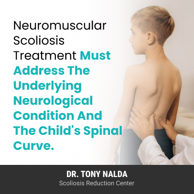
When there is a larger neuromuscular condition contributing to the spine’s unnatural position, the neurological condition needs to drive treatment planning, so a child’s curve, including its cause, needs to be fully understood.
An MRI for Atypical Curve Patterns
Most scoliosis curves are right-bending, and this helps protect the heart, so when an X-ray shows a left-bending curve pattern, an MRI may be recommended (2).
A left sided thoracic curve pattern is considered atypical and can be highly progressive, and an MRI can rule out if there is an issue with the structure of the spinal cord or brain stem where the brain connects to the spine.
The brain and spinal cord work in tandem as the central nervous system, so nerve-related issues can involve the spinal cord or nerve roots where they exit the spine.
An MRI for Rapid Curve Progression
An MRI may be recommended for cases of rapid curve progression to better understand if there are other factors driving the curve’s rapid progression.
Rapid progression may also indicate spinal cord involvement because it’s an atypical feature in some cases, and for curves that are increasing quickly and causing nerve-related symptoms, an MRI may be suitable (2).
An MRI scan allows radiologists to see what’s happening inside the spinal cord and its immediate surroundings including the nerves and soft tissues.
When a curve is progressing quickly, it may be moving the patient towards spinal fusion surgery because once a certain amount of progression has happened, the spine may be beyond nonsurgical correction and spinal fusion may be needed to stabilize the spine.
If there is an underlying neurological issue, it needs to be addressed before surgery, and in some cases, identifying and addressing a spinal cord issue early may support proactive curve management to help slow progression and avoid future surgical intervention.
When curves are assessed accurately from the outset and are fully understood and treated proactively, some patients can avoid surgical treatment. Spinal fusion surgery can have its own risks, side effects, and potential complications (3).
If you or a family member has recently been diagnosed with scoliosis, an early and thorough specialized assessment can help determine whether an MRI is needed, whether a curve should be recommended for monitoring, scoliosis-specific bracing, physical therapy, chiropractic care, or a surgical referral.
Conclusion
What patients and families should know about an MRI scan for scoliosis is that it’s a safe painless procedure that helps us better understand a patient’s curve for treatment planning.
There is a lot of information to process following a scoliosis diagnosis, but there is help available, and no one should be making treatment decisions without the guidance of an experienced scoliosis specialist,
Generally, an MRI isn’t needed to diagnose or monitor scoliosis, but in cases where a curve is atypical, has an abnormal pattern, neurological symptoms are present, or is progressing rapidly, an MRI analysis helps us see if the spinal cord is involved, or if there are issues with the brainstem that may impact nerve communication between the brain and spine.
A better understanding of a patient’s curve and situation supports informed guidance and treatment recommendations so patients and their families can make informed treatment decisions and be more involved in the process.
References:
- Li AW, Chang A, Murphy JS, Li Y, Roye B, Hardesty CK, Glotzbecker MP; Pediatric Spine Study Group. Current practices in MRI screening in early onset scoliosis. Spine Deform. 2025 May;13(3):961-966. doi: 10.1007/s43390-024-01033-4. Epub 2025 Jan 22. PMID: 39838245; PMCID: PMC12021731
- Barnes PD, Brody JD, Jaramillo D, Akbar JU, Emans JB. Atypical idiopathic scoliosis: MR imaging evaluation. Radiology. 1993 Jan;186(1):247-53. doi: 10.1148/radiology.186.1.8416573. PMID: 8416573
- Fan H, Wang Q, Huang Z, Sui W, Yang J, Deng Y, Yang J. Comparison of Functional Outcome and Quality of Life in Patients With Idiopathic Scoliosis Treated by Spinal Fusion. Medicine (Baltimore). 2016 May;95(19):e3289. doi: 10.1097/MD.0000000000003289. PMID: 27175629; PMCID: PMC4902471