

There have been a number of spinal-imaging advancements over the years. EOS imaging uses low dose biplanar radiography to capture images of the spine from all angles, simultaneously, for a clear reconstructed 3D image of the spine as one long structure. Imaging is needed to diagnose and monitor scoliosis, so some patients need frequent X-rays.
EOS imaging for scoliosis is low-dose X-ray imaging that provides 2D and 3D images of a patient’s skeletal structure. What patients and their families should know is that EOS imaging uses approximately 50 to 85 percent less radiation exposure so is safer for children, images of the spine should be taken while in a standing or sitting position by an experienced operator, and EOS captures full-body images.
Scoliosis curves need to be monitored closely because most will progress with growth and increasing age.
EOS Imaging System
EOS is a low-dose imaging system that minimizes radiation exposure while capturing high-quality images of the spine from all angles for a 3-dimensional understanding of its structure and alignment.
The underlying nature of scoliosis is structural as it involves the spine’s vertebrae curving unnaturally to the side and rotating, and as a progressive condition, most scoliosis curves will increase which is why all curves should be closely monitored.
Even curves initially diagnosed as mild can change, and progression makes the spine more rigid, and in most cases, less responsive to treatment, and for patients who progress across the surgical threshold, invasive spinal surgery may be recommended.
When curves are diagnosed early, monitored closely, and treated proactively, however, many scoliosis curves are responsive to nonsurgical care.
While signs of scoliosis can usually be detected with a physical examination and an adam’s forward bend test, the only way to diagnose scoliosis is to capture images of the spine itself. Imaging is an important factor, but a patient’s medical history, symptoms, and treatment goals are also considered.
To diagnose scoliosis, an X-ray has to confirm the spine’s unnatural curve to the side, that the size of the curve is more than 10 degrees, and that there is also vertebral rotation (spine’s bones are also twisting).
Low Radiation Dose
Because scoliosis is so often diagnosed in children, and because most childhood curves are progressive due to growth, some young patients may be recommended for frequent X-rays to monitor how the spine is responding to growth and/or treatment.
Children and adults develop scoliosis, but when growth stops, curve progression usually slows down or stops, so generally, adults aren’t exposed to as much frequent imaging as some young patients with progressive curves.
Using low-dose EOS over traditional X-rays helps minimize the risk of cell damage over time, and because children are still growing, they have more remaining years for long-term effects of early and repeated exposure to radiation to develop.

EOS imaging is also faster than traditional X-rays because the imaging system uses two pairs of X-ray tubes working from different angles at the same time and can scan the entire body in up to 30-45 seconds.
For monitoring scoliosis over time, EOS can include a micro-dose setting to lower radiation exposure further, and an EOS scan uses approximately 95-percent less radiation than traditional CT (computed tomography scans).
EOS imaging can put parents’ minds at ease about the amount of radiation their children are being exposed to over time during curve monitoring and care.
Body Position During EOS Imaging
Patients and parents should understand the importance of scoliosis X-ray results. During a patient’s initial scoliosis X-ray, a number of factors are considered and evaluated including curve size, location, amount of rotation, curve flexibility, and curve progression risk.
It’s important that scoliosis imaging is taken while the patient is in a standing or sitting position. This gives an accurate image of the spine while under the load effects of gravity, along with the patient’s posture and sagittal balance.
It’s essential that curve measurements are determined accurately, and assessing a patient’s curve thoroughly is not a quick and simple process. EOS system imaging requires a skilled operator to create a precise and accurate 3D reconstructed image of a patient’s spine and lower body.
A patient’s treatment planning is customized around the results of a scoliosis X-ray, and measurements such as the Cobb angle are referred back to during treatment to compare how the spine is responding to treating, growth, or increasing age.
Even curves assessed as stable because they aren’t increasing at the time can change quickly during growth or age-related spinal degeneration, and any increasing symptoms suggest progression and changing treatment needs.
A skilled EOS system operator knows how to position the patient, quickly scan the entire spine, accurately measure the curve, its rotation, flexibility, and more, and most importantly, knows how to interpret the results and frame them into a proactive treatment plan.
Full-Body Images
EOS imaging is also unique because it offers full-body images that don’t need to be stitched together for a single image.
Traditional X-rays require multiple images that computer software combines for a final image, but these images aren’t as clear as EOS capturing seamless frontal and side views of the spine, pelvis, and lower limbs in a single exposure.
Scoliosis describes the spine’s unnatural curve and twist, but its effects can be widespread and extend to other areas of the body. Pelvic obliquity and leg length discrepancies may develop, and these secondary scoliosis complications are easier to detect and evaluate when they are captured in a single image in relation to the spine.
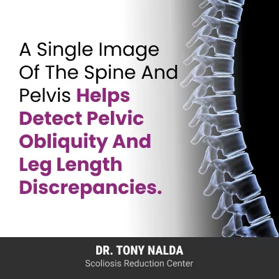
Having a single image of the spine, as it performs under gravity, its surroundings, and the entire body gives us a clear understanding of what’s happening in and around the spine for whole-body treatment planning.
Here at the Scoliosis Reduction Center®, we want to impact the spine’s structural nature first and foremost, but we also want to address the effects of scoliosis, and a primary effect is unbalanced posture that can contribute to uneven wear on the spine, its surrounding muscles, and supportive structures over the years.
Having a clearer image of a patient’s skeletal structure means having a clearer understanding of the scoliosis curve and its effects, and this supports more customized curve management for better patient outcomes in some cases.
Conclusion
EOS imaging is a fast and painless procedure that provides a single image of the spine from the front and sides so its structure, alignment, balance, and stability can be thoroughly assessed.
It also uses low-dose biplanar radiography so radiation exposure is limited, and this is significant particularly for young patients whose curves need to be closely monitored via X-ray during growth and treatment.
EOS imaging has the ability to create 3D reconstructions of the spine, and because scoliosis causes the spine to curve and twist unnaturally, it’s a 3D condition that needs to be monitored and addressed as such.
The dual X-ray bars capture images of the spine from different angles at the same time for faster procedure time, whole-body imaging, high precision when measuring curve size and alignment, and reducing the risk of cell damage due to repeated radiation exposure.
If you or a family member has been recently diagnosed with scoliosis, the progressive nature of scoliosis and its need for close monitoring should be considered, and for parents of young children concerned about the amount of radiation a child is exposed to during care, EOS imaging is an evidence-based alternative to traditional x rays.